
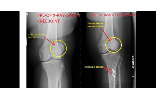
This is the case of a 36 year old female, who was seen in July of 2016. She has been having recurrent dislocation of her left knee for many years. This then progressed, becoming more severe with “locking” and mechanical symptoms of giving out. She has marked crepitus (gritty/grinding sensation) in the knee on activity. On clinical examination of her left knee by one of our experienced Orthopaedic Consultants at the Fracture and Orthopaedic Clinic, there is marked patellar apprehension(test for an abnormally positioned patella) with a range of motion of 0-120 degrees with no meniscal signs, no forward/backward laxity(loosening), and there was a mild effusion (swelling). Initial X-rays showed that this patient has significant patella alta (High riding patella) with lateral tracking (abnormal position of the patella).
An MRI was then requested which revealed that she has early patello-femoral osteo-arthritis, and also showed complete disruption of the medial patello-femoral ligament. Because of these issues, it was suggested to this patient that she have a procedure done called a “Distal Tubercle Transfer” to reduce her patella alta and bring her patella in line with the groove. She also requires a “Medial Patello-Femoral Ligament Reconstruction” for stabilization of her patella, to avoid further dislocation.
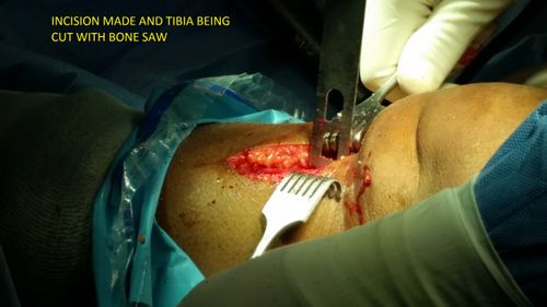
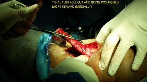
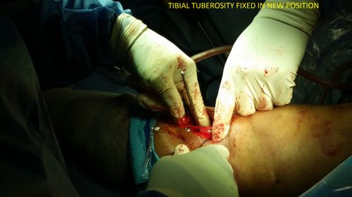
In October of 2016 this patient first had an arthroscopy (insertion of camera) of the knee, to visualise inside the joint before the major procedure was initiated ( The Distal Tubercle Transfer). The arthroscopy confirmed a high riding patella with erosion of cartilage down to bone. It also confirmed the presence of an extremely unstable patella that was sliding abnormally to the outer aspect of the joint. A distal femoral re-alignment was then performed. A piece of the shin bone that attaches to the patellar tendon was cut and moved to the inner aspect of the leg and fixed using screws, thus fixing the position of the patella back to normal. The position of the patella was checked during the surgery with an X-ray, which confirmed that the patella was back in its original position. Surgical cuts were then closed with sutures. Patient is yet to be followed up.

Posted:
Posted:
Posted:
Posted:
Posted: