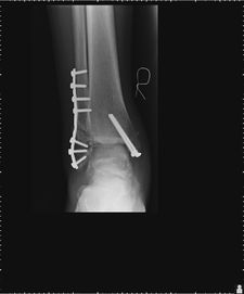
Mr. A.N., a 39 year old male labourer who injured his right ankle , was seen at another institution and was put in a cast and advised on the need for open reduction and internal fixation (insertion of plate and screws) of his injury. At the time of his arrival to the Fracture and Orthopedic Clinic, he was comfortable in his cast and had blisters on the medial(inner) aspect of the affected ankle. An X-ray was done which showed a displaced bi-malleolar fracture of his right ankle. Patient was advised on the need for surgery, and on the following day, he had an open reduction and internal fixation performed by Mr. Araujo of the F.A.O.C. The fibular fracture was cleaned and reduced, and an 8-hole plate was applied in reduction and held with screws. The medial malleolar fracture was then cleaned and reduced and held with a provisional K-wire and two compression screws. He was then advised to bear no weight on the right ankle for six weeks, after which he could only be allowed to partial weight bear thereafter.
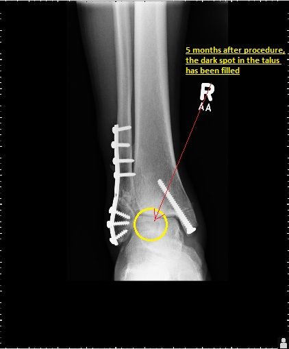
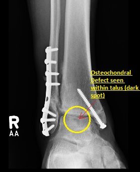
Mr A.N. Was relatively fine and progressing very well until at about 6 months post operatively, he began to get pain and swelling during his physiotherapy, which later progressed and got worse. He was sent for a CT scan of his right ankle at seven months post op, for suspicion of an intra-articular OCD (Osteochondral Defect). The CT scan performed did confirm the presence of an Osteochondral defect (OCD) of his right talus and he was advised on the need for a second surgery.
Mr, A.N's second surgery consisted of viewing of the right ankle joint with a scope (arthroscopy) with debridement and insertion of bone tissue from a donor,(allograft) into the osteochondral defect of his talus. The bone tissue was sourced from a Tissue Bank in the United States. One week after his second surgery, all surgical cuts were healing nicely, there were no signs of infection and Mr. A.N. Stated he experienced no pain in his ankle. He was advised to do gentle exercises of his right ankle and not to weight bear for six weeks, to allow the allograft to "take" and incorporate itself nicely into his talar bone. About two months after his second surgery, Mr. A.N. Was fully weight bearing on his right ankle with minimal discomfort and NO PAIN. At four months post op, he continued to improve and his X-rays at that point showed healing of the grafted area in the talus. Five months post op , he was discharged from our clinic, and given clearance to return to work with normal duties (apart from climbing, due to mildly limited range of motion of his right ankle) with an open appointment to see again if any problems arise.
Posted:
Posted: